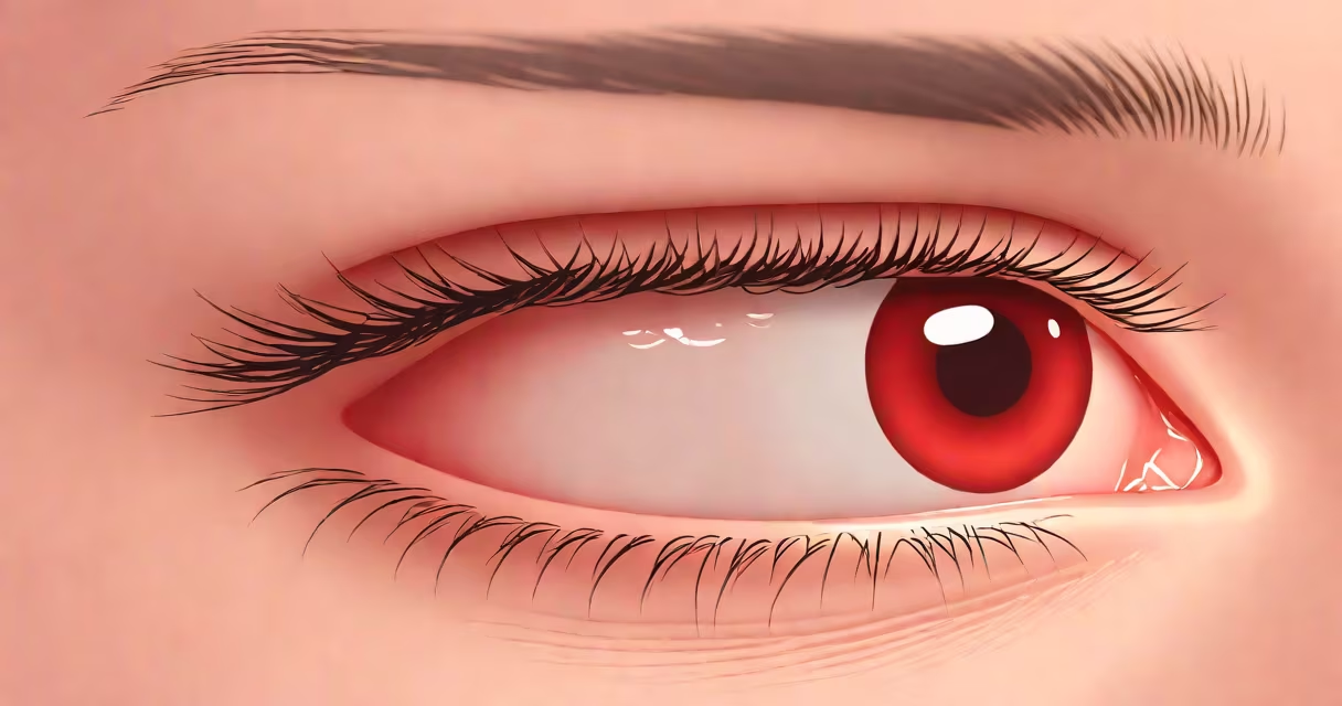
A swollen tear duct occurs when the nasolacrimal duct, which drains tears from the eye into the nose, becomes blocked or inflamed. This blockage leads to fluid buildup, swelling near the inner corner of the eye, excessive tearing, and sometimes pain or discharge.
The condition often stems from congenital blockages in infants, infections like dacryocystitis in adults, or chronic inflammation. It may also result from trauma, tumors, or age-related narrowing of the duct. Early recognition helps prevent complications such as abscess formation or permanent scarring.
Swelling typically appears at the junction between the eye and nose, just below the inner eyelid. If left untreated, persistent blockage can lead to recurrent infections and impaired vision due to constant moisture on the cornea.
Tears are produced by the lacrimal glands and drain through small openings called puncta at the inner corners of the eyelids. From there, they flow through the canaliculi into the lacrimal sac and then down the nasolacrimal duct into the nasal cavity. When any part of this pathway is obstructed, tears cannot exit properly, leading to backup and swelling.
Blockages most commonly occur at the nasolacrimal duct's narrowest point-the valve of Hasner-where congenital membrane remnants may persist in babies. In adults, obstruction often arises from chronic inflammation, scar tissue from prior infections, or structural changes due to aging. A single episode of infection can cause temporary swelling, but recurring episodes suggest an underlying anatomical issue.
For example, a 2018 study published in *Ophthalmology* found that 62% of adult patients with chronic epiphora (excessive tearing) had confirmed nasolacrimal duct obstruction via dacryoscintigraphy, a nuclear imaging test that tracks tear flow.
Swollen tear ducts fall into two broad categories: congenital and acquired. Congenital cases affect newborns and young children, while acquired cases develop later in life and are more frequently linked to infection or structural changes.
| Type | Typical Age Group | Primary Cause | Key Symptom |
|---|---|---|---|
| Congenital | Born to 2 years old | Membrane at valve of Hasner fails to open | Persistent tearing, mucus discharge from eye |
| Acquired (infection) | Adults, especially 40+ | Dacryocystitis from bacteria like Staphylococcus aureus | Redness, swelling, pain, pus drainage |
| Acquired (non-infectious) | Older adults, particularly women | Scarring, tumors, autoimmune disease (e.g., sarcoidosis) | Gradual onset of tearing, no pain initially |
One non-obvious distinction: many assume swelling near the eye is due to allergies or conjunctivitis. But true tear duct swelling involves localized pressure and tenderness at the medial canthus-the inner corner of the eye-rather than diffuse redness or itching. Allergies cause watery eyes but not the firm, painful lump associated with a blocked duct.
Another common confusion is equating a swollen tear duct with a stye. While both can appear as lumps near the eyelid, a stye forms along the lash follicle and resolves within days. A swollen tear duct persists longer, worsens with pressure, and may require medical intervention.
Not all swelling goes away on its own. If swelling lasts more than 1-2 weeks, especially with pus, fever, or worsening pain, consult an ophthalmologist. Delayed treatment increases risk of orbital cellulitis-a serious infection spreading to surrounding tissues.
I once saw a patient who ignored mild swelling for six weeks. By the time he presented, his entire upper eyelid was swollen, and he had a temperature of 101.3°F. Imaging revealed a large abscess in the lacrimal sac. He required IV antibiotics and surgical drainage. This outcome is preventable with early evaluation.
Diagnosis begins with a physical exam and probing of the puncta. If blockage is suspected, a dye test (fluorescein nasolacrimal irrigation) confirms whether fluid flows normally. For persistent cases, CT or MRI scans detect tumors or structural abnormalities.
Preventing blockages involves treating sinus infections promptly and avoiding eye rubbing, which can introduce bacteria into the duct system. For infants, gentle massage of the lacrimal sac area (from inner corner toward nose) may help open a congenital blockage. Studies show this technique resolves blockage in 70% of cases within the first year.
For adults, managing dry eyes or chronic sinusitis reduces strain on the tear drainage system. Avoid using expired eye drops or sharing makeup-both increase infection risk.
When conservative measures fail, procedures like dacryocystorhinostomy (DCR)-a surgery that creates a new drainage route from the lacrimal sac to the nasal cavity-are effective. Success rates exceed 90%, according to the American Academy of Ophthalmology.
Understanding what causes a swollen tear duct enables timely action. Persistent tearing, pain, or visible swelling near the eye should prompt medical evaluation. Early diagnosis prevents complications and restores normal tear function.