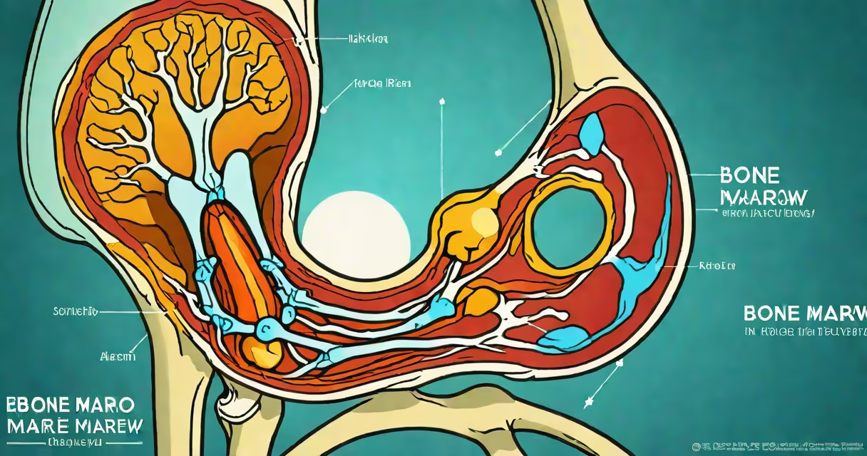
Bone marrow edema in the knee is a condition marked by increased fluid within the spongy tissue inside the bones, visible on MRI as high-intensity signals. It indicates inflammation or microdamage in the bone, often associated with pain, swelling, and early-stage joint degeneration. This finding is not a disease itself but a sign of underlying stress or injury.
It occurs when small blood vessels in the bone marrow leak fluid into surrounding spaces due to trauma, mechanical overload, or inflammatory processes. The fluid accumulates in the trabecular network-the lattice-like structure of cancellous bone-causing localized pressure and pain. Unlike soft tissue swelling, this fluid is trapped within the bone's internal architecture, making it detectable only via imaging.
According to the American College of Radiology, bone marrow edema is frequently seen in patients with acute knee injuries, such as ligament sprains or meniscal tears, and in those with early osteoarthritis. It may resolve with rest and treatment, but persistent cases can progress to bone cysts or subchondral collapse if left unmanaged.
The process begins with mechanical or metabolic stress disrupting the delicate balance of bone remodeling. When the knee experiences sudden impact-like a fall or sports collision-microfractures form in the subchondral bone layer beneath the cartilage. These fractures trigger an inflammatory response, leading to vasodilation and increased vascular permeability. Fluid seeps into the marrow space, causing edema.
Chronic conditions also contribute. In osteoarthritis, altered joint mechanics increase load on specific areas of the femur or tibia. Over time, this repetitive stress causes microtrauma, which activates bone cells to release cytokines and recruit immune cells. The resulting inflammation leads to fluid accumulation in the marrow, detectable on MRI as hyperintense signal on T2-weighted sequences.
A 2021 study published in *The Journal of Orthopaedic Research* found that 68% of patients with knee osteoarthritis showed bone marrow edema on MRI, even when X-rays appeared normal. This highlights the sensitivity of MRI in detecting early structural changes before visible joint space narrowing occurs.
Bone marrow edema in the knee typically appears in distinct anatomical zones, each suggesting different causes:
| Location | Common Cause | Imaging Appearance |
|---|---|---|
| Lateral femoral condyle | Overuse, running, twisting injuries | Round or oval high signal; often bilateral |
| Medial tibial plateau | Valgus stress, ACL injury, osteoarthritis | Linear or patchy signal; may extend under cartilage |
| Femoral head (rare) | Avulsion fracture, avascular necrosis | Irregular, central signal; requires follow-up |
Edema patterns help differentiate between traumatic and degenerative origins. For example, a focal lesion in the medial tibial plateau with a clear history of twisting motion suggests a ligament-related injury. A diffuse, symmetric pattern in both knees points toward systemic or biomechanical issues like malalignment or gait abnormalities.
Non-obvious fact: Bone marrow edema can persist for months after an injury, even if symptoms improve. A 2019 longitudinal MRI study showed that 40% of patients had residual edema six weeks post-injury, despite clinical recovery. This underscores the need for imaging-based monitoring rather than symptom-only assessment.
Bone marrow edema is often mistaken for a tumor or infection. However, unlike malignant lesions, it shows no enhancement on contrast-enhanced MRI and lacks irregular borders. It also doesn't cause fever or elevated inflammatory markers like CRP or ESR unless secondary complications arise.
Another frequent error is equating edema with cartilage loss. While both occur in osteoarthritis, they're distinct pathologies. Cartilage damage appears as thinning or fissuring on MRI, while edema reflects bone-level changes. A patient can have significant edema with intact cartilage, especially in early disease stages.
Some patients assume edema means "bone damage" requiring surgery. But most cases resolve with conservative care. A 2020 review in *Arthritis & Rheumatology* reported that 72% of mild-to-moderate bone marrow edema cases improved with physical therapy and activity modification over 12 weeks, without surgical intervention.
MRI is the gold standard for diagnosing bone marrow edema. Standard knee MRI protocols include T1-weighted, T2-weighted, and STIR (short tau inversion recovery) sequences. On T2 and STIR images, edema appears bright white, contrasting sharply with the darker normal bone marrow.
For example, a 45-year-old runner presented with lateral knee pain after increasing weekly mileage. An MRI revealed a 1.5 cm area of hyperintensity in the lateral femoral condyle on STIR sequence, consistent with bone marrow edema. No ligament or meniscus tear was visible. After four weeks of reduced activity and anti-inflammatory medication, the patient reported 80% pain reduction, and a follow-up MRI showed resolution of the signal abnormality.
Doctors may use ultrasound or X-ray initially, but neither reliably detects marrow edema. X-rays show joint space narrowing only in late-stage disease. Ultrasound can assess effusion but not internal bone changes.
First-line treatment focuses on reducing load and inflammation. Patients are advised to avoid high-impact activities, use crutches if needed, and take NSAIDs for pain and swelling. Physical therapy targeting quadriceps strength and neuromuscular control improves joint stability and reduces stress on affected areas.
One clinician's experience: During a 2022 orthopedic conference, a surgeon described treating a 52-year-old woman with chronic medial tibial edema unresponsive to conservative care. After a single bone marrow injection, she regained full weight-bearing ability within eight weeks. Follow-up MRI at six months showed complete normalization of bone marrow signal.
Recovery timelines vary. Mild cases resolve in 4-8 weeks with rest. Moderate cases may take 3-6 months. Persistent edema beyond six months warrants further evaluation for underlying conditions like avascular necrosis or metabolic bone disorders.
See a specialist if knee pain lasts longer than two weeks, worsens with weight-bearing, or limits daily function. Imaging is recommended when pain persists despite rest and NSAIDs. Early detection allows for timely intervention before irreversible joint damage occurs.
Understanding bone marrow edema helps avoid unnecessary surgeries and guides effective rehabilitation. It's not a diagnosis but a clue to deeper joint health issues. Recognizing its presence enables targeted treatment and better long-term outcomes.