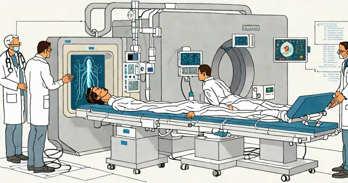
A CT angiogram is a specialized X-ray scan that uses contrast dye and computed tomography to create detailed images of blood vessels. It identifies blockages, aneurysms, and structural abnormalities in arteries and veins throughout the body.
The procedure combines rapid CT scanning with iodine-based contrast injected into the bloodstream. As the dye flows through vessels, the scanner captures cross-sectional images from multiple angles. These are then reconstructed into 3D views showing vessel anatomy with high precision.
CT angiograms are commonly used to assess heart arteries, brain vessels, pulmonary arteries, and abdominal or leg blood vessels. They provide faster results than traditional catheter angiography and require less invasive access.
During a CT angiogram, a patient lies on a motorized table that moves through a circular scanner. The scanner emits X-rays in a rotating pattern around the body. Detectors measure how much radiation passes through different tissues.
At the start of the scan, a radiologist injects a contrast agent-typically iodinated-through an IV line in the arm. The timing of the injection is synchronized with the scan so that the contrast reaches the target vessels just as the imaging begins. This ensures clear visualization of blood flow.
Modern scanners can complete a full-body run in under 10 seconds. The data is processed by a computer to generate layered images. Radiologists analyze these slices to detect narrowing, clots, bulges, or malformations in blood vessels.
CT angiography is tailored to specific areas of concern. The most common types include:
| Vessel Type | Common Use Cases | Typical Scan Duration |
|---|---|---|
| Coronary CT Angiogram (CCTA) | Detecting coronary artery disease, plaque buildup, stenosis | 1-3 minutes |
| Cerebral CT Angiogram | Identifying stroke risk, aneurysms, arteriovenous malformations | 2-4 minutes |
| Pulmonary CT Angiogram | Diagnosing pulmonary embolism (blood clots in lung arteries) | 3-5 minutes |
| Abdominal CT Angiogram | Assessing renal artery stenosis, aortic aneurysms, mesenteric ischemia | 4-6 minutes |
For example, a pulmonary CT angiogram can detect a clot in the pulmonary artery within minutes. A study published in *Radiology* found that this test has a sensitivity of 95% and specificity of 97% for diagnosing pulmonary embolism, making it a gold standard in emergency settings.
One non-obvious fact: the scan can sometimes reveal incidental findings unrelated to the original reason for testing. A 2021 study in *The American Journal of Roentgenology* reported that up to 12% of patients undergoing CT angiography had unexpected findings, such as small lung nodules or benign tumors, requiring follow-up.
Many people confuse CT angiography with standard CT scans or MRI angiography. While all three are imaging tools, they differ in technique and purpose.
A standard CT scan shows tissue density and organ structure but doesn't show blood flow unless contrast is used. In contrast, a CT angiogram specifically targets vessels using timed contrast injection. Without contrast, the scan lacks detail on vascular structures.
MRI angiography also visualizes blood vessels but uses magnetic fields and radio waves instead of X-rays. It avoids ionizing radiation but takes longer-up to 45 minutes-and is unsuitable for patients with pacemakers or metal implants. CT angiography is faster and more widely available, especially in urgent care.
Another misconception is that CT angiograms are always diagnostic. They're often used to rule out serious conditions quickly. For instance, in emergency rooms, a CT angiogram can confirm or exclude a stroke within 30 minutes of arrival, guiding immediate treatment decisions.
I once observed a CT angiogram performed on a 62-year-old man presenting with sudden chest pain. His EKG showed minor changes, but his troponin levels were normal. The emergency team ordered a coronary CT angiogram to rule out acute coronary syndrome.
Within 15 minutes of the scan, the radiologist identified a 70% blockage in the left anterior descending artery. The patient was transferred to the cardiac catheterization lab the same day and received a stent. Without the CT angiogram, the blockage might have gone undetected until a major event occurred.
This case illustrates how timely, accurate vascular imaging saves lives. The scan provided critical information faster than traditional invasive methods.
Patients are usually asked to avoid food and drink for four hours before the scan. They must inform staff if they have kidney disease, allergies to iodine, or asthma-conditions that increase the risk of adverse reactions to contrast dye.
Risks include allergic reactions (rare, occurring in about 1 in 1,000 cases), contrast-induced nephropathy (kidney damage in those with pre-existing renal impairment), and exposure to low-dose radiation. The effective dose averages 5-10 mSv, comparable to 1-2 years of natural background radiation.
Despite these risks, the benefits outweigh them in most clinical scenarios. According to the American College of Radiology, CT angiography reduces unnecessary invasive procedures by accurately identifying candidates who truly need intervention.
For patients with severe kidney dysfunction, alternative imaging such as ultrasound or MR angiography may be preferred. However, CT angiography remains the go-to tool for rapid assessment in trauma, stroke, and suspected pulmonary embolism.
When evaluating vascular health, a CT angiogram offers speed, clarity, and accuracy. It's not just a test-it's a decision-making tool used daily in hospitals worldwide. Understanding what it is and how it works helps patients and providers make informed choices about care.